Hello. I'm Dr. Dongkyu Lee, an orthopedic specialist. If you've experienced shoulder calcific tendinitis, you know the pain can be extremely severe. Even when the pain isn't at its worst, persistent discomfort and difficulty with daily activities can significantly reduce your quality of life. That's why patients visit the hospital seeking treatment. Unfortunately, most hospitals only offer pain injections and extracorporeal shockwave therapy. Of course, depending on the condition, these treatments alone can sometimes be sufficient. However, when the calcific deposit is large, these treatments (injections, shockwave) aren't effective enough, and patients are told they need surgery. From my perspective, this is truly unfortunate. The fundamental treatment for calcific tendinitis is removing the calcific deposit. Conservatively, pain injections are given first to manage symptoms, and extracorporeal shockwave therapy is used to try to eliminate the deposit. However, shockwave therapy alone rarely removes the calcification completely. As a result, patients get injections and feel better temporarily, only to have the pain recur, then get more injections and improve briefly before relapsing again... This cycle repeats, with recurring pain affecting both activities and daily life, drastically reducing quality of life. After this cycle continues, patients are eventually told it's time for surgery. For patients who can't have surgery or don't want it, hearing that surgery is the only option left is truly disheartening. Many patients also visit the hospital and are told they need surgery right away because their calcific deposit is too large. Surgery for calcific tendinitis involves removing the calcification from within the tendon, which means cutting through the tendon to access and remove the deposit. Arthroscopic surgery is commonly performed, and a device called a shaver is used to remove the deposit. While it would be ideal to remove only the calcification, the deposit infiltrates between the tendon fibers, so healthy tendon tissue inevitably gets damaged as well. Furthermore, the larger the calcific deposit, the larger the incision in the tendon needs to be, resulting in greater tendon damage. One patient had such a large deposit that during surgery, the surgeon removed only a partial amount to minimize tendon damage, leaving residual calcification that continued to cause pain. That patient then came to me. After receiving the non-surgical "calcific deposit aspiration" procedure, this patient was freed from pain. Shoulder calcific tendinitis can be treated non-surgically. It can be treated with calcific deposit aspiration. Let me show you a case. The patient was a woman in her 50s who had suffered from calcific tendinitis for 2 years. She had received injections whenever the pain flared up and had undergone dozens of shockwave treatments. However, the pain kept recurring, and she was eventually told she needed surgery, which is when she came to see me. She traveled from another region, and at the time of her visit, the pain was so severe she couldn't even move her arm.
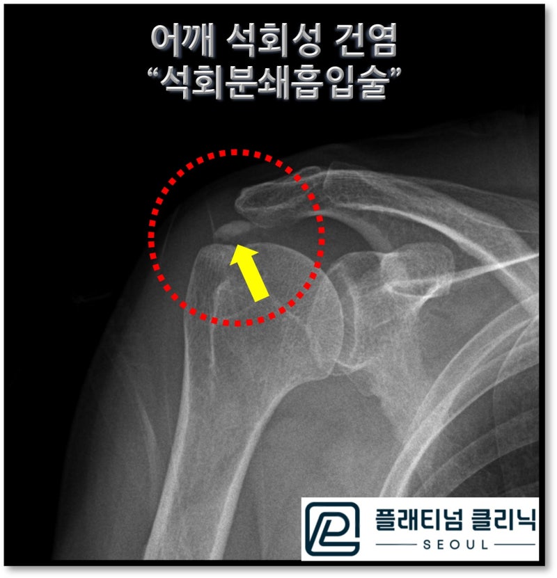
On the X-ray, the white area indicated by the yellow arrow within the red circle is the calcific deposit. A considerably large deposit can be observed. To address the root cause, calcific deposit aspiration was performed.
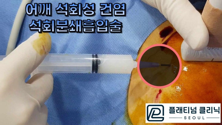
This is the actual procedure video of the calcific deposit aspiration. At first, you can see white, chunk-like calcification being extracted. After crushing the deposit, clean saline solution is used to repeatedly flush and aspirate the area, and you can observe the clear water turning cloudy from the dissolved calcification.
During the actual procedure, ultrasound guidance is used. If you look carefully at the ultrasound footage, you can see the deposit area expanding and deflating repeatedly. Initially, the interior appears white and cloudy, but toward the end, the interior becomes clean as it expands and deflates, meaning all removable calcification has been successfully extracted.
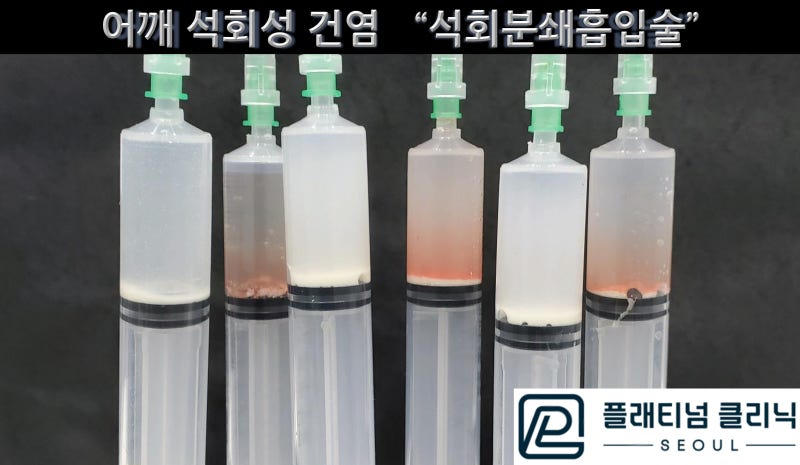
The process shown in the video involves using multiple syringes in a repetitive manner. Looking at the syringe photo after the procedure, you can see a large amount of white calcification settled at the bottom of the syringe.
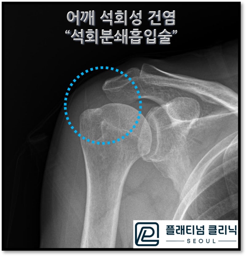
The X-ray taken after the procedure shows that the calcific deposit visible before treatment has disappeared.
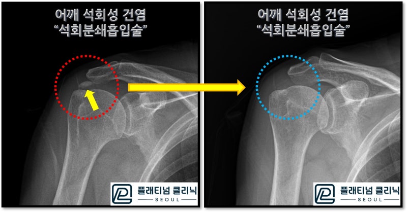
Comparing the before and after images side by side, the removal of the calcific deposit is even more clearly evident. When this patient first visited, she couldn't even move her arm due to pain. After the procedure, she was smiling, able to move her arm, and said the pain had decreased dramatically. When I showed her the before and after images and the syringe photo proving the deposit was gone, she expressed deep gratitude, saying she wished she had received this treatment sooner. I will continue working hard until every patient suffering from shoulder calcific tendinitis can be cured without surgery.
Shoulder calcific tendinitis can be fundamentally treated without surgery. Calcific Deposit Aspiration