Hello. I am Dr. Dongkyu Lee, an orthopedic specialist.
Calcific tendinitis can be treated without surgery.
https://blog.naver.com/9690067/222801103857
🔗 https://blog.naver.com/9690067/222801103857We receive many inquiries from patients across the country. "Where can I get percutaneous calcium deposit aspiration?" "Why isn't this treatment available where I live?" "My hospital says the deposit is too large and surgery is needed -- is it really possible to treat without surgery?" These are among the many questions we receive. Percutaneous calcium deposit aspiration is quite a demanding procedure. The calcium must be precisely targeted under ultrasound guidance. Depending on the nature of the calcium, very hard deposits can be extremely difficult to break up. Very sticky calcium can clog the syringe, making aspiration difficult. The degree of fragmentation must also be judged in real-time under ultrasound, as excessive fragmentation can damage the tendon. When calcific tendinitis is accompanied by a tear, aspiration becomes more challenging, requiring 2-way or 3-way techniques to remove the calcium. All of this requires experience and skill. Without them, percutaneous calcium deposit aspiration cannot be easily performed. That is why some patients come to us after unsuccessful procedures at other hospitals. Some hospitals only treat calcific tendinitis surgically. Today's case is also a patient who came from a distant area and was told the calcium was too large for anything but surgery. This patient had suffered for years with calcific tendinitis, repeatedly getting injections and shockwave therapy only to have the pain return. This time the pain became so severe that even injections no longer helped, and they were told at a large hospital that surgery was necessary.
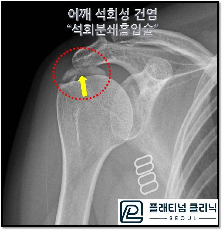
The X-ray shows an enormous amount of calcium deposit indicated by the arrow within the red circle. The patient had already brought an MRI from another hospital.
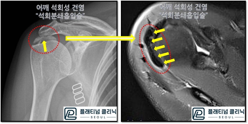
The MRI on the right shows a massive calcium deposit (black area indicated by yellow arrows) near the infraspinatus, measuring nearly 4-5cm. With such a large deposit, it is understandable that most hospitals would recommend surgery. Some doctors may not even know about non-surgical calcium removal. Others may know that fragmenting and aspirating such a large deposit is challenging and simply find it easier to recommend surgery. We performed percutaneous calcium deposit aspiration non-surgically.
The video shows the guide being precisely inserted into the calcium under ultrasound, followed by fragmentation using a specialized tip. Saline solution is then injected to wash out and aspirate the calcium. This particular patient's procedure took approximately 15-20 minutes.
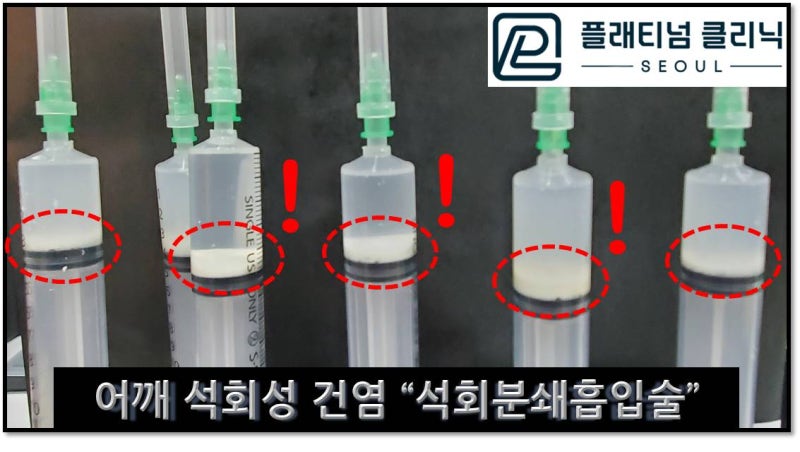
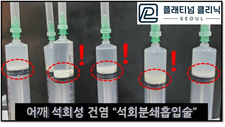
The post-procedure syringe photo shows white calcium settled at the bottom. The total amount aspirated from all syringes combined was approximately 3-4cc.
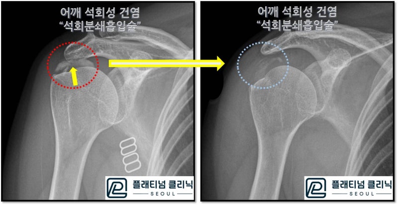
The post-procedure X-ray shows the calcium in the red circle (pre-procedure) has been almost completely absorbed in the blue circle (post-procedure). The remaining small amount of calcium will either be absorbed naturally or can be accelerated with additional shockwave therapy. After the local anesthesia wore off, the patient said the pain that was present before the procedure had almost completely disappeared. The patient returned to their hometown to receive additional shockwave therapy there.
As you can see, a large calcium deposit does not mean surgery is the only option. Even large deposits can be properly fragmented and removed. Calcific tendinitis can be sufficiently treated non-surgically.