When treatment is needed for calcific tendinitis of the shoulder, tendon injury, or muscle tear, hospitals often recommend X-ray, ultrasound, or MRI. Some patients believe MRI provides the most accurate results. However, MRI is not always necessary when treating calcific tendinitis, frozen shoulder, rotator cuff tears, or other shoulder pain. So when and how should you get properly diagnosed? Recently, a patient came to our clinic after repeated treatment for a rotator cuff tear without improvement. At a previous hospital, MRI showed a partial rotator cuff tear, and the patient had received 7-8 rounds of expensive regenerative injections with no improvement. Let's first examine this patient's X-ray and MRI.

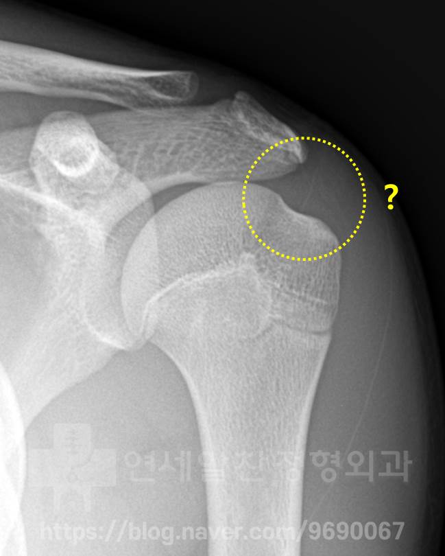
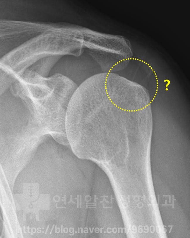
The X-ray shows completely normal, unremarkable findings.
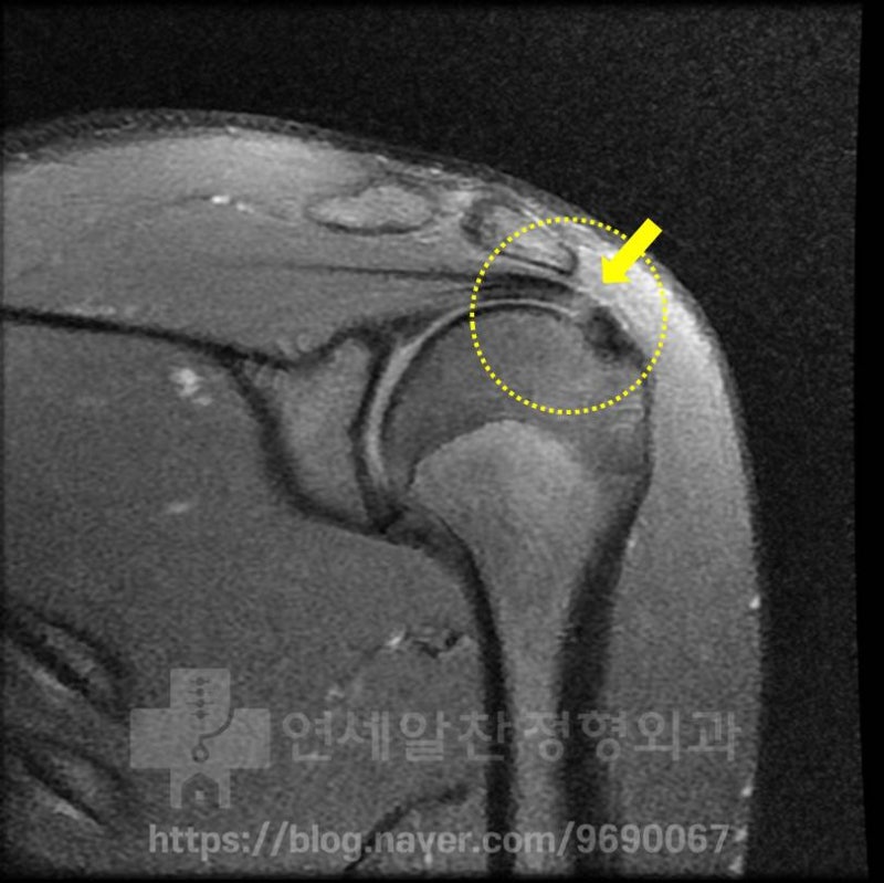
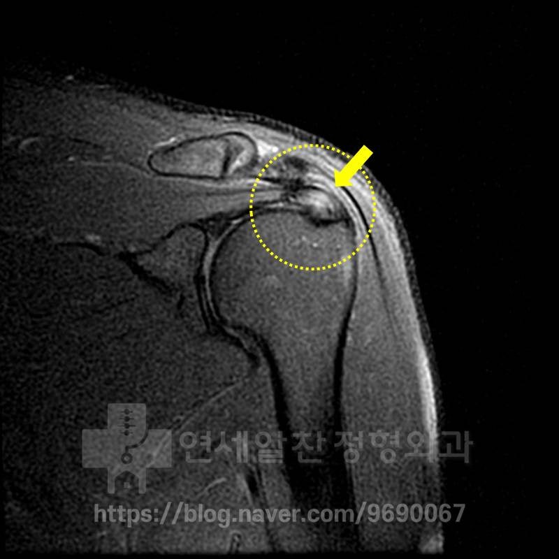
On MRI, the supraspinatus area within the yellow dotted line shows a slightly whitish appearance where the arrow points. This indicates some inflammation within the supraspinatus, which was diagnosed as a partial tear leading to regenerative injection treatment. However, based on this MRI finding, it appears to be simple inflammation without progression to actual tearing. Since some time had passed, we decided to re-examine with ultrasound.
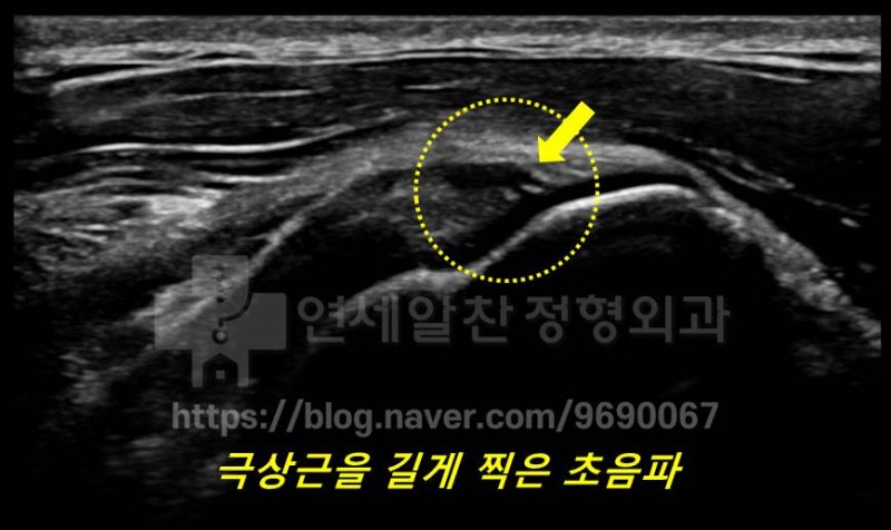
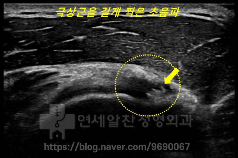
On this ultrasound image, the supraspinatus is shown in long axis. Within the yellow dotted area, the arrows point to about 2 white dots, which are calcifications. These are tiny micro-calcifications less than 1mm, and the surrounding ligament appears dark, indicating inflammation. Normally, the ligament should appear white on ultrasound; the dark appearance around it means inflammation is present.
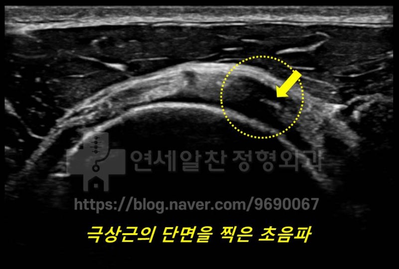
This ultrasound image shows the supraspinatus in cross-section. Similar to the previous image, calcification is visible where the arrows point within the yellow dotted area, with surrounding darkening. In this patient's case, severe inflammation of the supraspinatus was caused by micro-calcifications. After approximately 5 sessions of shockwave therapy, the symptoms improved. Micro-calcifications like these are often invisible on X-ray, and since MRI scans at 4mm intervals, calcifications smaller than 4mm may be missed. With ultrasound, because the examiner moves the transducer and scans the entire ligament in real time, if the ultrasound resolution is good and the examiner is experienced, calcific tendinitis can be detected via ultrasound rather than MRI, enabling targeted treatment. Let me share another patient case. This patient was told on MRI that the ligament was torn and surgery was recommended. The patient came to our clinic to get a second opinion before proceeding with surgery. Let's examine the X-ray and MRI.
The X-ray shows normal findings.
On MRI, the supraspinatus ligament appears white instead of the normal black, showing severe inflammation. This was interpreted as severe tendinosis (ligament weakening), and surgical repair was recommended. However, upon closer inspection, a small dark spot can be seen within the supraspinatus on MRI, suggesting micro-calcification. So we decided to confirm with ultrasound.
On ultrasound, 3 white dots are visible where the arrows point within the yellow dotted area, with surrounding darkening. This indicates very severe inflammation caused by micro-calcifications. This patient also improved after 7 sessions of shockwave therapy. Most calcific tendinitis of reasonable size can be diagnosed with X-ray alone. However, small micro-calcifications may not be visible on X-ray. Many people think MRI is necessary for treating calcific tendinitis, but since MRI scans at 4mm intervals, calcifications smaller than 4mm can be missed. With ultrasound, experienced physicians can detect even small micro-calcifications. Ultrasound can also distinguish the nature of calcifications (hard calcifications in the formative phase vs. cheese-like calcifications in the resorptive phase), making it a very useful diagnostic tool for determining the treatment direction.