Hello. I'm Dr. Dongkyu Lee, an orthopedic specialist. Today I'll show you a case of calcific tendinitis treated non-surgically with calcific deposit aspiration. For information about calcific deposit aspiration, please refer to the link below. https://blog.naver.com/9690067/222801103857
🔗 https://blog.naver.com/9690067/222801103857This case involves a patient who had suffered from calcific tendinitis for 2 years. The patient had only received injections when the pain flared up and underwent shockwave therapy, but the pain kept recurring and the patient was in great distress. After 2 years of this recurring pain, the patient couldn't sleep well, their daily routine fell apart, and their quality of life had deteriorated severely. Let me get straight to the case.
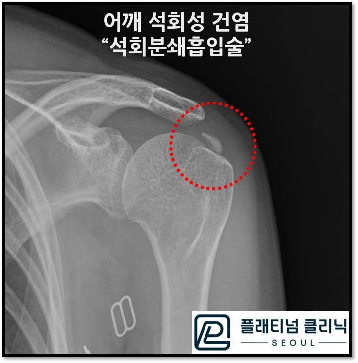
As shown on the X-ray, calcification is visible above the humerus within the red circle.
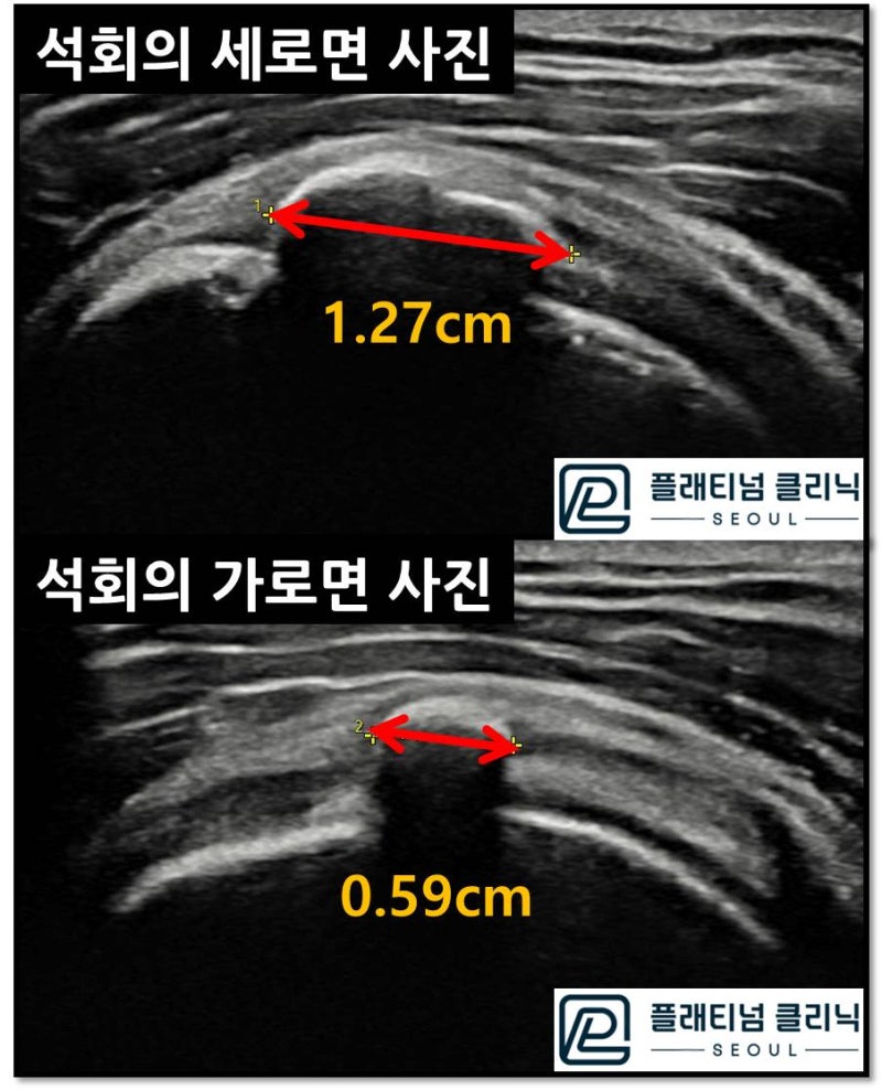
Ultrasound was used to check the size and nature of the calcification. The deposit measures 1.27 cm in length and 0.59 cm in width. The dark acoustic shadow below the deposit on ultrasound indicates a hard, solid calcification. Ultrasound cannot penetrate solid materials, so bone appears as a dark shadow below, and since the area below this deposit also appears dark like bone, it indicates a hard calcification. Calcific deposit aspiration was performed to free the patient from 2 years of suffering in one session.
In the video above, you can see that the initially clear syringe gradually fills with white calcification as the aspiration continues, until the syringe is completely white at the end. After crushing the calcification, saline solution is used to flush and aspirate the fragments.
During the procedure, ultrasound guidance is used. In the video, you can see the bulging and deflating within the red circle. This is the process of crushing the calcification interior, then repeatedly injecting and aspirating saline solution to remove the deposits. Once the calcification is sufficiently crushed, most of it can be removed.
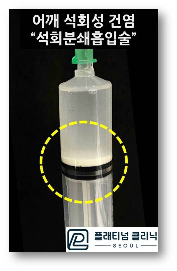
After the calcific deposit aspiration, when the syringe is left standing upright for a while, you can see the calcification settle to the bottom as shown in the photo. Once the calcification is removed this way, the fundamentally eliminated deposit also significantly reduces the pressure and inflammation within the tendon, and the agonizing pain disappears. One of the things patients struggle with most is being unable to sleep due to pain. After the procedure, many patients tell me they finally had a comfortable night's sleep for the first time in ages and express their gratitude.
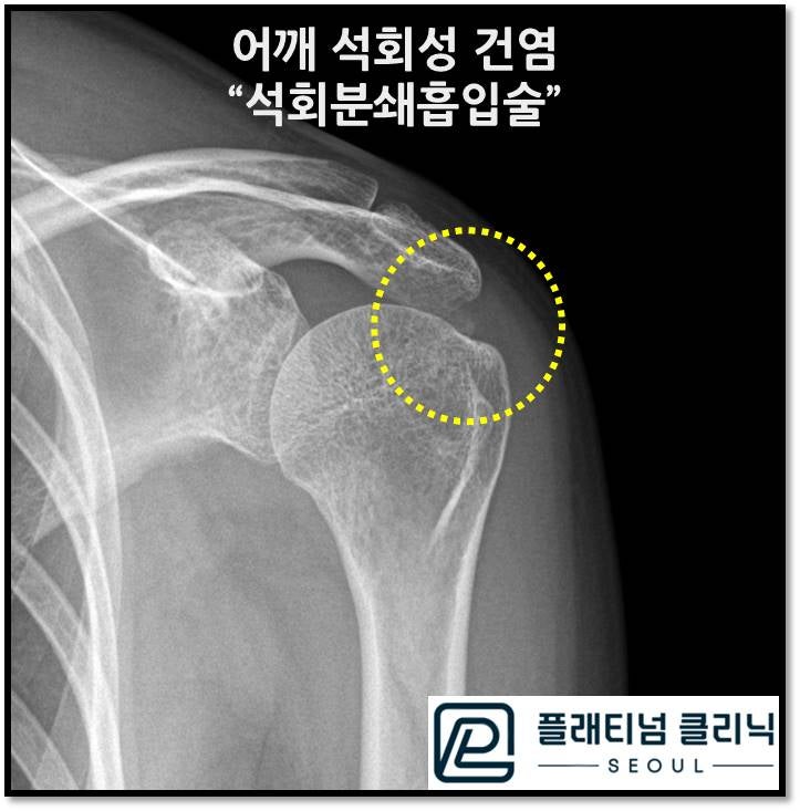
After the procedure, the X-ray shows that the calcification visible initially is nearly gone. The small amount of remaining calcification can naturally absorb on its own or be eliminated through additional extracorporeal shockwave therapy.
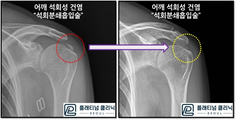
Comparing the before and after images, the removal of the calcification is even more clearly evident. So many patients suffer from calcific tendinitis. Because the pain is significant, quality of life deteriorates and patients truly struggle.
Calcific tendinitis can be cured non-surgically. Furthermore, treatment is possible in a single session. (Additional injection and shockwave therapy may be needed.)