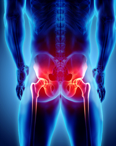
Our body has many types of joints — shoulder, knee, spine, hip, and more. In clinical practice, the joints most commonly treated are mobile (synovial) joints. Among these, the hip joint has a relatively stable structure and doesn't commonly develop problems. However, when issues do arise — from extensive athletic activity, car accidents, or falls — the hip can be particularly difficult to treat. Calcific tendinitis primarily occurs in the shoulder and is widely known as such. However, it is not exclusively a shoulder condition — calcium can deposit in any joint where tendons pass. It can occur in the hip, elbow, wrist, ankle, and any other joint in the body.

Calcific tendinitis occurs when calcified material deposits in tendons, hardens, and then causes pain as it dissolves in each respective joint. When calcium forms in the hip, the pain can be so severe that walking normally becomes impossible, potentially being confused with leg or spinal disc conditions — making prompt and accurate diagnosis essential. Hip calcific tendinitis symptoms include persistent pain in the buttocks and pelvis, potentially extending to the thigh, with severe night pain and inability to sit cross-legged or perform external rotation combined with abduction. Like shoulder calcific tendinitis, hip calcific tendinitis progresses through a hardening phase lasting months to years, followed by a dissolution phase lasting weeks to months. Through the formation, maintenance, and absorption phases, calcium may naturally disappear. However, natural resolution can take years, and when hardened calcium begins to dissolve, it causes extreme hip pain requiring treatment. Similar to shoulder calcific tendinitis, the formation phase doesn't cause severe pain, but symptoms appear mainly during the dissolution phase — or calcium may remain dormant before causing severe pain later. This dissolution-related pain release is sometimes called a 'chemical abscess.'

Hip calcific tendinitis can be diagnosed with a simple X-ray, which shows calcified areas as white spots. Initial treatment involves medication for pain and inflammation control, along with extracorporeal shockwave therapy to promote blood circulation in the tendons around the hip. Beyond treatment, daily management is crucial — proper exercise intensity and rest are essential. If increased exercise leads to more hip pain, it's best to rest for about two weeks while focusing on hospital treatment. If improvement doesn't occur after treatment, additional imaging with ultrasound or MRI should be performed to accurately diagnose the affected area and check for additional conditions. Theoretically, high-impact sports like soccer or running are not recommended for hip calcific tendinitis. It's safest to consult your doctor and resume exercise only when daily pain has essentially resolved.