Hello. I'm Dr. Dongkyu Lee, an orthopedic specialist. Shoulder calcific tendinitis most commonly occurs in the supraspinatus but can develop in any tendon. When calcific tendinitis occurs in the supraspinatus, it's easily visible on frontal X-rays and can be readily diagnosed. However, when it occurs in other locations, X-rays being two-dimensional images means the calcium deposit can be obscured by bone and potentially missed. In particular, when calcific tendinitis is in the subscapularis, the calcium is located at the front of the shoulder and overlaps with bone on X-ray, which can lead to missed diagnoses. This patient had been examined at another hospital but was told it was just inflammation. After treatment without improvement and with excruciating pain, the patient came to me.
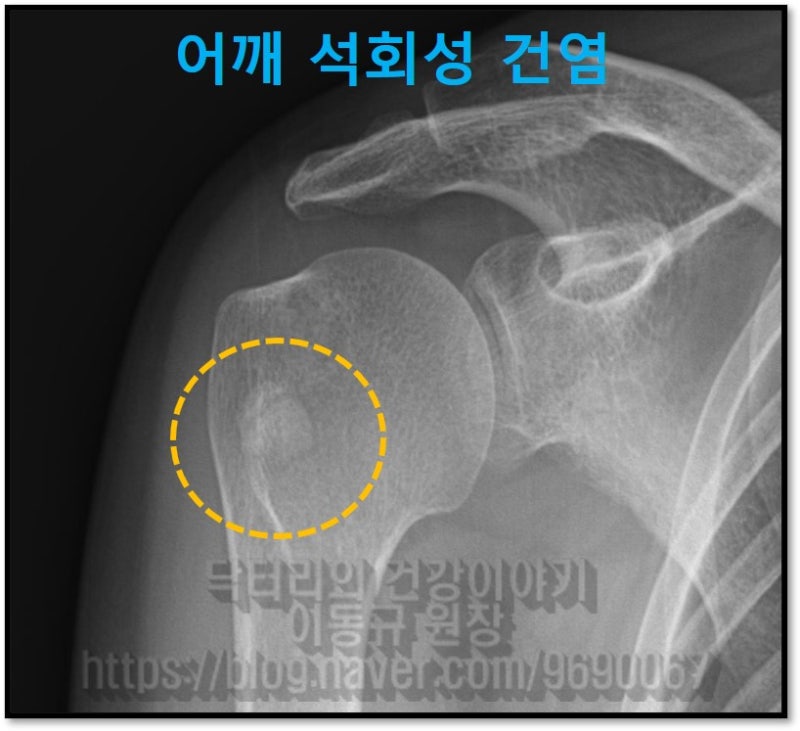
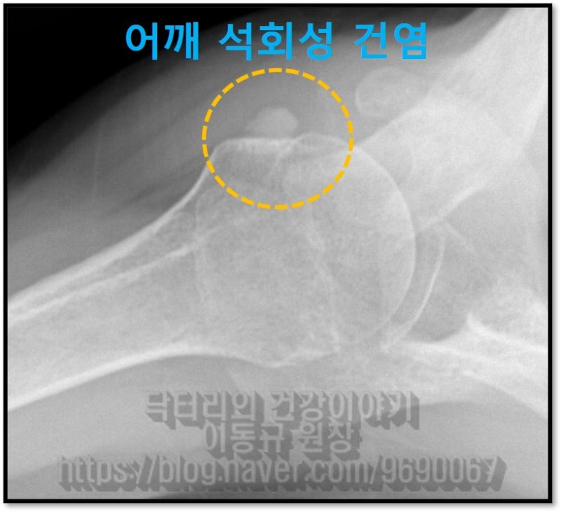
Had the X-ray been examined more carefully, or had the patient's symptoms been observed more closely, accurate diagnosis would have been possible.
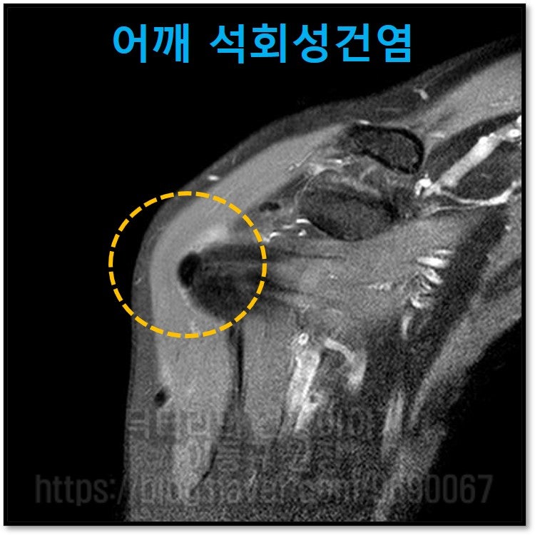
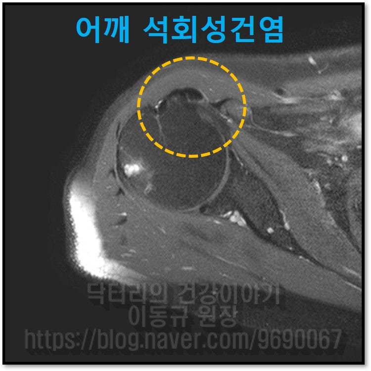
On MRI, the dark area within the yellow circle corresponds to calcific tendinitis.
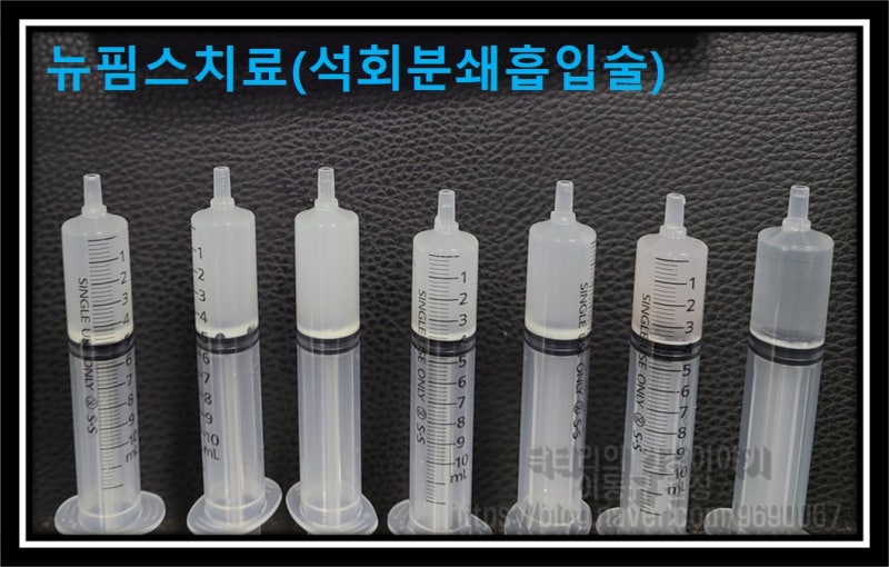
Due to the patient's extreme shoulder pain, NewPIMS treatment (calcium crushing and aspiration) was performed immediately to remove the calcium.

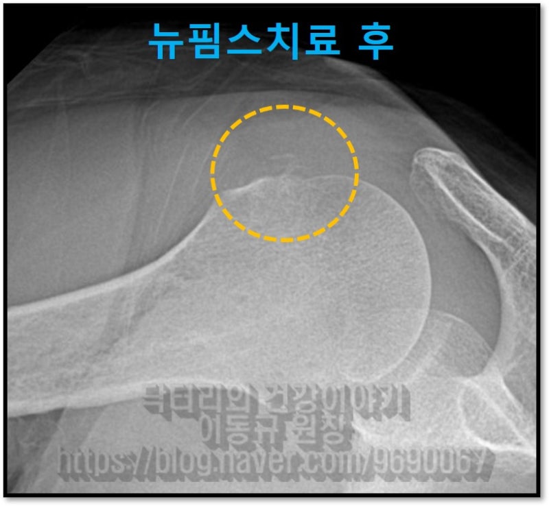
Post-treatment X-ray shows that the calcium visible before the procedure has been nearly completely removed.
The patient experienced immediate symptom improvement after the procedure. Shoulder calcific tendinitis causes extreme pain due to increased pressure and inflammation from the calcium deposit. Once the calcium is removed, the pressure and inflammation immediately subside, resulting in rapid pain relief. The patient had been receiving ineffective treatment and was very satisfied to finally receive proper treatment that resolved the pain. As with any condition, accurate diagnosis is essential first, and once a diagnosis is made, appropriate treatment should follow. Furthermore, receiving treatment specialized for the specific condition produces the best results. For shoulder calcific tendinitis, NewPIMS treatment (calcium crushing and aspiration) is the most targeted treatment available.